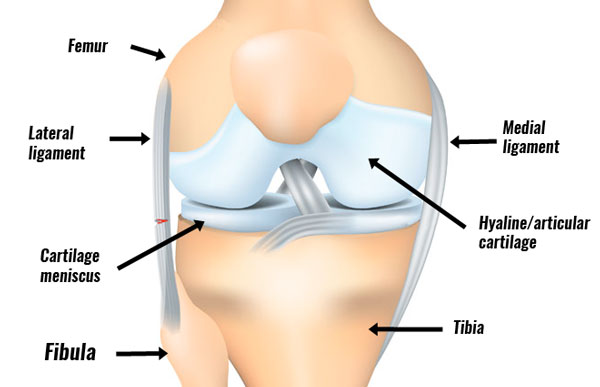
An LCL sprain or lateral knee ligament sprain is a tear to the ligament on the outside of the knee. It most commonly occurs following a direct blow to the inside of the knee. However, it can also develop gradually through overuse. Our full LCL rehab program takes you step-by-step from initial injury to full fitness.
Medically reviewed by Dr Chaminda Goonetilleke, 13th Dec. 2021
Symptoms of an LCL sprain
The main symptoms of a lateral collateral ligament sprain (LCL sprain) are:
- Pain on the outside of the knee.
- Symptoms vary from being very mild to a complete rupture of the ligament.
- You may have swelling over the outside of the joint, especially with more severe injuries.
Lateral ligament sprains are categorized into grades 1, 2, or 3 depending on the extent of your injury.
Grade 1 LCL sprain
If you have a grade 1 sprain your knee will feel tender on the outside. You will have little or no swelling. However, you will feel pain with the varus stress test (see below), but no joint laxity.
Grade 2 LCL sprain
With a grade 2 LCL sprain, you will have significant tenderness on the outside of your knee. You will likely have some swelling. The varus stress test will indicate pain and some joint laxity. However, you will have a definite endpoint that indicates your ligament is still intact.
Grade 3 LCL sprain
A grade 3 lateral ligament sprain is a complete tear of the ligament. Pain can vary and may be actually less than a grade 2 sprain. You will have significant joint laxity with the varus stress test, with no firm endpoint. Your knee may be very unstable.
A full examination is needed once any pain and swelling have gone down. A painful swollen knee is more difficult to assess.
Varus stress test
The varus stress test is used to help diagnose lateral knee ligament sprains. It stresses the lateral ligament specifically.
Your doctor/physio holds your leg with the knee slightly bent to approx 30 degrees. They stabilize your thigh whilst applying inward pressure on your lower leg.
As a result, the lateral ligament is stretched/stressed. The test is positive if you feel pain on the outside of your knee. The degree of damage can be determined by how much movement/instability is present.
Imaging
In more serious cases an MRI scan and/or X-Ray may be necessary.
Anatomy

The lateral collateral knee ligament or LCL for short connects the femur (thigh bone) to the top of the fibula (shin bone). The ligament itself is a narrow strong cord of collagen fibres and its function is to provide stability to the outside of the knee.
The ligament is not connected to the lateral meniscus in the joint like the medial ligament (on the inside) does. Therefore, LCL sprains are not normally associated with cartilage meniscus tears. However, injury to the anterior cruciate ligament or posterior cruciate ligaments can occur at the same time as an LCL sprain.
Causes
The LCL is most commonly injured by a direct impact on the inner surface of the knee. For example, in a rugby or a football tackle.
Force on the inside of the knee causes the joint to open on the outside, therefore stretching the lateral ligament. An LCL sprain is less common than a medial collateral ligament sprain.
Treatment
Treatment consists of immediate first aid PRICE principles (Protection, Rest, Ice, Compression, Elevation). This is followed by a full rehabilitation program. Treatment methods include:
- Cold therapy & compression
- Wearing a knee support or brace
- Knee ligament taping
- Massage
- Rehabilitation exercises
- Surgery (not always required)
